About HIFU for Prostate Cancer

Making a decision about your prostate cancer treatment can be very confusing. The good news is, if you are focused on the biochemical disease-free rate – the PSA level after treatment –  most therapy options are nearly equal.

Many men, however, also worry about their quality of life after the prostate cancer procedure. In consultation we often hear questions such as “Will I regain the same erection as before the treatment?” and “Will I have total control of bladder after the procedure?” This is where the choice of prostate cancer treatment option matters the most because the results vary so widely depending on the procedure.

When treating a patient for prostate cancer, we have three objectives – the “trifecta” – on which we base the success of the procedure:

  • Cancer control: PSA levels
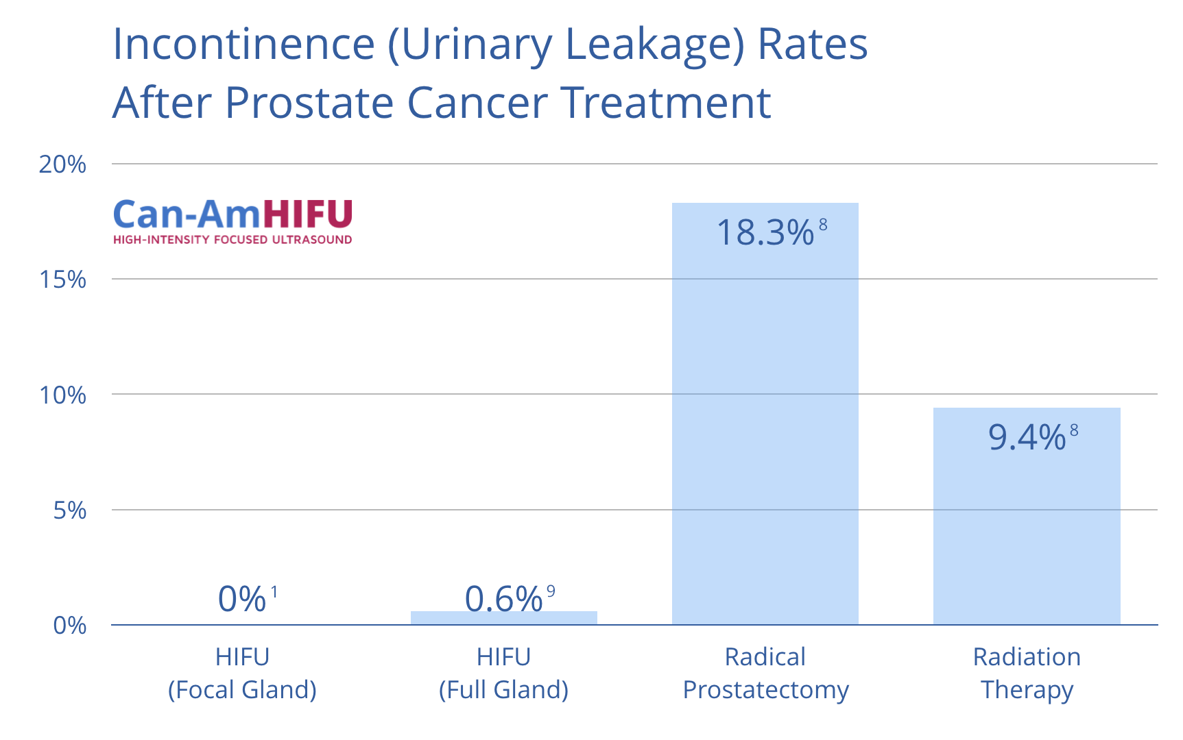
  • Continence: normal urinary function
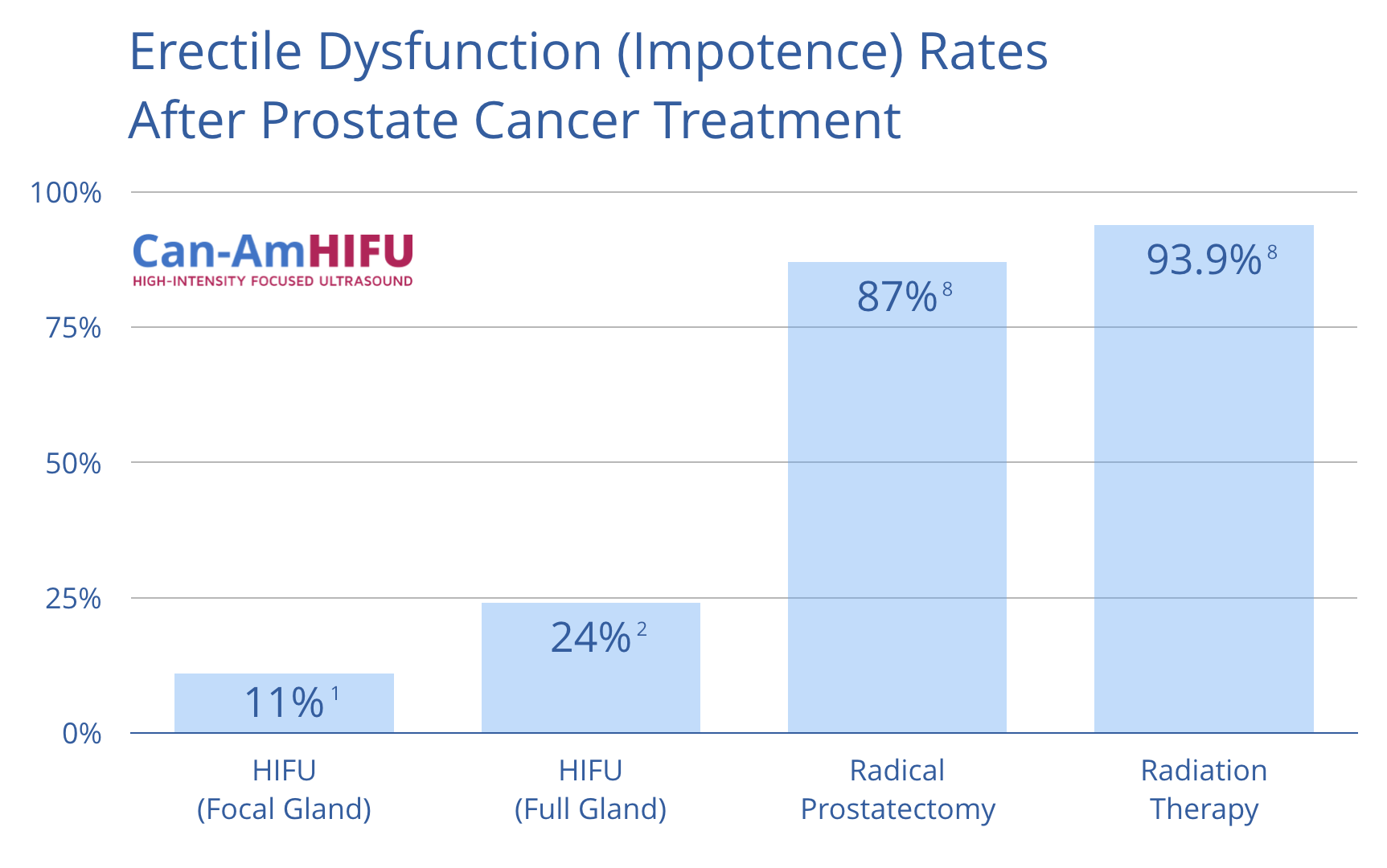
  • Potency: preservation of erectile function

Sonablate HIFU is precise and flexible, allowing our physicians to customize the treatment plan to each patient’s prostate cancer diagnosis with the goal of achieving these key objectives. Below is a summary of the different levels of treatment customization and their clinical results.

Standard (Full Gland) HIFU Procedure

During a standard Sonablate HIFU procedure, referred to as “total ablation”, we treat the entire prostate gland. This option is most appropriate for patients who have organ-confined prostate cancer that has spread to multiple areas of the prostate.

The clinical results of a standard HIFU procedure:

  • 92% of patients are successfully treated5
  • Over 99% of men remain continent5
  • 70% of men retain erectile function5
Focal HIFU Therapy

Focal HIFU is becoming common for the treatment of localized prostate cancer. With focal HIFU therapy, only the actual tumour sites are treated instead of the whole prostate gland. This is ideal for patients that have fewer than 4 cores positive on their biopsies, particularly if all of the positive cores are in the same area of the prostate.

Prior to recommending a focal treatment, we will verify the patient’s biopsy results with an MRI scan. We use the MRI results to confirm that the only areas affected are the ones shown by the biopsy. If MRI indicates additional suspicious areas, we then suggest an “MRI-Ultrasound Fusion”-targeted biopsy of those areas. This approach greatly reduces the chances of leaving prostate cancer behind.

Focal HIFU is intended to help avoid the important structures located near the prostate and reduce the risk of side-effects such as erectile dysfunction. Focal therapy may be suitable for men with low- or intermediate-risk cancer who have a small tumor volume on one side of the prostate or a few small tumor sites even if on both sides of the gland.

Focal HIFU allows for a shorter procedure, usually 20-90 minutes, and a quicker return to normal life and an even lower risk of side-effects.

The clinical results of a focal HIFU procedure:

  • 95% maintained potency (defined as return of erections sufficient for penetrative sex)6
  • 89% of men achieved the trifecta status (defined as pad-free, leak-free continence, erections sufficient for intercourse and cancer control at 12 months)6
Salvage Treatment with HIFU

Sonablate HIFU may be performed on patients who have previously been treated for prostate cancer and their cancer has returned. This form of treatment is often referred to as a “salvage treatment”. A salvage HIFU treatment can be performed on patients who have been treated with radiation, cryotherapy or previous HIFU procedure, where there is biopsy proof of recurrence and tissue that can be visualized with the ultrasound. A clinician will have to determine if the patient is a candidate for salvage HIFU.

If a patient has already undergone a focal HIFU treatment and we identify new sites of prostate cancer, we can then either repeat a focal treatment or recommend total ablation with HIFU.

Clinical outcomes of salvage Sonablate HIFU following external beam radiation therapy (EBRT):

  • 71% biochemical disease free rate7
  • 93% of men remain continent7

HIFU vs Other Therapies: Sexual Function and Continence After Prostate Cancer Treatment

Erectile Dysfunction (Impotence) Rates After Prostate Cancer Treatment

Incontinence (Urinary Leakage) Rates After Prostate Cancer Treatment


Here’s what you can expect before, during and after a HIFU Procedure.
Before the Treatment

One day before the HIFU, you must only consume clear fluids. You will then have to purchase two enemas. On the day of the treatment, you will self-administer them before coming to the clinic. The second enema should be taken after the first one has cleared. You will need to empty your bowel two hours before you head in for the procedure.

When you arrive at our clinic, the nurse will greet you and prepare you for your procedure. To keep you still during your treatment, you will be given either a) a spinal anesthetic with light IV sedation, or b) a general anesthesia. The choice of anesthetic will depend on your medical history and whether you are taking anti-coagulants:

  • If you recently started taking an anti-coagulant, in most cases we will recommend general anesthesia;
  • If you have been taking an anti-coagulant for a while, you may be able to stop taking the medication for 5 days prior to HIFU. In this case, we can proceed with the spinal anesthetic and IV sedation.
  • If you are not taking anti-coagulants, you will receive spinal anesthetic and IV sedation.

Regardless of the type of anesthetic, you will continue breathing on your own throughout the procedure. Your HIFU doctor will discuss these options with you in advance.

During the Treatment

You will not feel any pain during the treatment. The small treatment probe is inserted through the rectum. The doctor uses this probe to capture real-time images of the prostate and the surrounding tissue. After studying these images, the same probe delivers the ultrasound waves directly into the prostate tissue. The treatment takes from one to three hours, depending on the amount of tissue being treated.

After the procedure is complete, we expect some swelling of the prostate as a reaction to the treatment. You will wear a catheter while this swelling goes down, which usually takes between 5 days to 3 weeks. This way your bladder can empty completely while your prostate is healing and the destroyed tissue is transported out of your body. You can resume all other aspects of your normal lifestyle, such as diet, returning to work, and recreational activities in just a few days after treatment. You will be sent home from the clinic a few hours after the treatment, and you will have no incisions to care for.

After the Treatment

You will stay at our clinic for one to two hours immediately after the procedure while the anesthetic wears off. Then you will be released to recover at your hotel or at home. You will take a mild antibiotic as prescribed by your physician. Your body may feel a little sore, but this will not last and is a normal result of any procedure performed under anesthetic. You may also have mild problems with your urinary system for the first two weeks after a HIFU procedure. These are also normal complaints and will resolve themselves as your prostate heals. Your doctor may prescribe some medication to speed up the healing.

High-Intensity Focused Ultrasound is a technology with over 40 years of research behind it. It is currently approved for prostate cancer treatment in over 50 countries, including Canada and the US.
Technology

The idea for HIFU began at the Indiana University School of Medicine in Indianapolis in the 1970s. Since that time, research centers worldwide have helped to perfect the design and make HIFU for the treatment of prostate cancer ready for global use.

To understand how the HIFU process works to destroy cancer cells, it is best to start with remembering something you likely did as a child with a magnifying glass. If you recall, when you focused the light from the sun onto a leaf on the ground at a certain angle, the leaf would burn.

The reason the leaf burned is because the magnifying glass causes the sun’s rays to focus at a point below the glass. This point of focus creates extremely high temperatures. You could have put the leaf in any other section of the light beam and it would not have burned. It is only at the point of focus there is enough heat to burn the leaf.

This is the same principle used in HIFU technology. The transducer replaces the magnifying glass, and the sound waves replace the rays from the sun. But the rest of the comparison remains very much the same.

During a HIFU treatment, the doctor will use real-time images to guide the transducer towards the prostate. The sound waves are then directed to intersect in the center of the prostate, where the temperature increases, thus destroying the cells. The real-time images are important to the success of the treatment, because they allow the doctor to make changes throughout the procedure for maximum effectiveness.

How It Works

The Sonablate uses a fully integrated ultrasound probe that handles both the imaging and the treatment of the prostate. Real-time imaging is key in creating a customized plan, especially in case of the focal HIFU. Once the treatment plan has been mapped, the doctor directs the machine to send a HIFU beam to the prostate cancer targets. As the treatment progresses, the doctor can compare these real-time images to the “before treatment” images on the same screen. This allows the doctor to adjust if necessary, which will help to ensure a successful treatment.

Research

Sonablate HIFU for prostate cancer has been extensively studied for over a decade. The following publications and presentations highlight clinical results on the use of the high-intensity focused ultrasound and specifically the Sonablate system which we use at Can-Am HIFU.

Focal Therapy for Localised Unifocal and Multifocal Prostate Cancer: a Prospective Development Study. Ahmed, Hashim U, Richard G Hindley, Louise Dickinson, Alex Freeman, Alex P Kirkham, Mahua Sahu, Rebecca Scott, Clare Allen, Jan Van der Meulen, and Mark Emberton. The Lancet Oncology 13, no. 6 (June 2012): 622–632.

Focal Therapy for Localized Prostate Cancer: A Phase I/II Trial. Ahmed, H.U., A. Freeman, A. Kirkham, M. Sahu, R. Scott, C. Allen, J. Van der Meulen, and M. Emberton. The Journal of Urology 185, no. 4 (April 2011): 1246–1255.

Transrectal High-intensity Focused Ultrasound for the Treatment of Localized Prostate Cancer: Eight-year Experience. Uchida, Toyoaki, Sunao Shoji, Mayura Nakano, Satoko Hongo, Masahiro Nitta, Akiko Murota, and Yoshihiro Nagata. International Journal of Urology 16, no. 11 (2009): 881–886.

High-intensity-focused Ultrasound in the Treatment of Primary Prostate Cancer: The First UK Series. Ahmed, H. U., E. Zacharakis, T. Dudderidge, J. N. Armitage, R. Scott, J. Calleary, R. Illing, et al. British Journal of Cancer 101, no. 1 (2009): 19–26.

Transrectal High-intensity Focused Ultrasound for Treatment of Localized Prostate Cancer. Inoue, Yoji, Keisuke Goto, Tetsutaro Hayashi, and Mutsuo Hayashi. International Journal of Urology 18, no. 5 (2011): 358–363.

Visually Directed Transrectal High Intensity Focused Ultrasound for the Treatment of Prostate Cancer: a Preliminary Report on the Italian Experience. Mearini, Luigi, Leonardo D’Urso, Devis Collura, Alessandro Zucchi, Elisabetta Costantini, Andrea Formiconi, Vittorio Bini, Giovanni Muto, and Massimo Porena. The Journal of Urology 181, no. 1 (January 2009): 105–111; discussion 111–112.

Visually directed high-intensity focused ultrasound for organ-confined prostate cancer: a proposed standard for the conduct of therapy. Rowland O. Illing*†, Tom A. Leslie‡, James E. Kennedy‡, John G. Calleary*, Christopher W. Ogden§ and Mark Emberton*† *The Institute of Urology and Nephrology, University College London, London, †The Clinical Effectiveness Unit, The Royal College of Surgeons of England, London, ‡Department of Urology, The Churchill Hospital, Oxford, and §Department of Urology, Chelsea and Westminster Hospital, London, UK Accepted for publication in British Journal of Urology 27 July 2006.

Five Year Experience of transrectal high-intensity focused ultrasound using the Sonablate device in the treatment of localized Prostate cancer. Toyoaki Uchida, Hiroshi ohkusa, Hideyuki Yamashita, Sunao Shojl, Yochihiro Nagata, Toru Hyodo and Takefumi Satoh. International Journal of Urology, Vol. 13, 228-233. 2006.

Outcome Analysis of High Intensity Focused Ultrasound for clinically localized prostate cancer in Japan- Seven-Year Follow-Up. Makoto Suzuki, M.D., Toyoaki Uchida, M.D., Toshiro Terachi, M.D..: Tokyo, Japan. (Presentation made by Dr. Suzuki at Western Sectional AUA Meeting in Maui, June 2006) Visually Directed HIFU for Prostate Cancer –a new standard. Rowland O. Illing**, Sam Dawkins*, Chris W. Ogden* and Mark Emberton**. *Institute of Urology and Nephrology, University College Hospital, London, UK ** The Clinical Effectiveness Unit, The Royal College of Surgeons of England, London, UK

Treatment of localized prostate cancer using high-intensity focused ultrasound. Toyoaki Uchida. Dept. of Urology University of Tokai Hachioji Hospital. British Journal of Urology International 2006.

Prostate Cancer Therapy with High-Intensity Focused Ultrasound -Comprehensive Review Thomas A. Gardner and Michael A Koch, Indiana University Medical Center, Indianapolis. Clinical Genitourinary Cancer Vol 4. No.3, 2005.

Transrectal High-Intensity Focused Ultrasound in the treatment of Localized Prostate Cancer: A Multicenter Study. Acta Urol. Jpn. 51 651-658, 2005. Toyoaki Uchida. The Dept. of Urology, Tokai University Hachioji Hospital.

High Intensity Focused Ultrasound for the Treatment of Localized Prostate Cancer. A Multi-Center Experience. George M Suarez*, Miami, FL; Rafael Estrella, Santiago De los Caballeros, Dominican Republic; Carlos Garcia, Puerto Vallarta, Mexico. Abstract from Presentation given at The 15th International Prostate Cancer Conference in Vail, Colorado, February 2005

Transrectal HIFU: The Next Generation? Prostate Cancer Research Institute (PCRI) Insights. Douglas O. Chinn, MD Chinn & Chinn Urology Medical Associates, Arcadia, CA February 2005. Pages 8-15.

Clinical Outcome of HIFU for the Treatment of localized prostate cancer: 5-year Experience. Toyoaki Uchida, Hiroshi Ohkusa, Hideyuki Yamashita, Yoshihiro Nagata Department of Urology, Tokai University Hachioji Hospital. Abstract – ISTU 2004

Clinical Outcome of HIFU for Localized Prostate Cancer: 5 year Observations. Toyoaki Uchida, E. Yamashita, Y. Okusa, T. Nagata, Tokai University, Tokyo, Japan. Podium presentation given at the 2004 Japan Endourology and ESWL Conference, Okayama, Japan November, 2004.

High Intensity Focused Ultrasound for Prostate Cancer: Clinical Results and Technological Evolution John C. Rewcastle, Ph.D. Department of Radiology, University of Calgary Alberta, Canada Quality of Life in Patients with High Intensity Focused Ultrasound (HIFU) for localized Prostate Cancer. Toyoaki Uchida, E. Yamashita, Y. Okusa, T. Nagata, Tokai University, Tokyo, Japan. Podium presentation given at the 2004 Japan Endourology and ESWL Conference, Okayama, Japan November, 2004.

Transrectal High-Intensity Focused Ultrasound for treatment of patients with Stage Tib-2N0M0 localized Prostate Cancer: A Preliminary Report. Presentation of preliminary clinical results of transrectal HIFU in stage T1b-2N0M0 prostate cancer. Dr. T Uchida, M.D.

Localized Prostate Cancer Treatment by High Intensity Focused Ultrasound (HIFU): Preliminary Results. Toyoaki Uchida M.D., Department of Urology, Kitasato University, Tokyo, Japan. This summary has been abstracted from previously published work and supplemented by additional new treatment data prepared by Dr. T. Uchida, M.D. by Focus Surgery, Inc.

Noninvasive Surgery of Prostate Tissue by High Intensity Focused Ultrasound: An Updated Report. European Journal of Ultrasound. N. T. Sanghvi 2,*, R. S. Foster1, R. Bihrle1, R. Casey3, T. Uchida4, M. H. Phillips2, J. Syrus2, and A. V. Zaitsev2 , K. W. Marich2 , F. J. Fry

1. Ahmed, HU. (2012). The Lancet Oncology. Focal Therapy for Localized Unifocal and Multifocal Prostate Cancer: A Prospective Development Study, 2045(12), 70121-3.

2. Uchida, T. (2012). 11th International Symposium on Therapeutic Ultrasound. AIP Conf. Proc. Twelve Years’ Experience with High-Intensity Focused Ultrasound (HIFU) Using Sonablate Devices for the Treatment of Localized Prostate Cancer. 1481, 401-406.

3. Novara, G. (2012). European Urology. Systematic Review and Meta-analysis of Studies Reporting Oncologic Outcome After Robot-assisted radical8Prostatectomy. 62, 382-404.

4. Alicikus, Z. (2011). Cancer. Ten-Year Outcomes of High-Dose Intensity-Modulated Radiotherapy for Localized Prostate Cancer. 1429-1437.

5. Ahmed, HU. (2009). BJC. High-intensity-focused ultrasound in the treatment of primary prostate cancer: the first UK series, 101, 19-26.

6. Ahmed, HU. (2011). Journal of Urology. Focal Therapy for Localized Prostate Cancer: A Phase I/II Trial, 185(4),1254-5.

7. Zacharakis, E. (2008). BJUI. The feasibility and safety of high-intensity focused ultrasound as salvage therapy for recurrent prostate cancer following external beam radiotherapy,102, 786-792

8. Resnick, M. (2013). The New England Journal of Medicine. Long-Term Functional Outcomes after Treatment for Localized Prostate Cancer. 368, 436-45.

9. Ahmed, HU. (2009). BJC. High-intensity-focused ultrasound in the treatment of primary prostate cancer: the first UK series, 101, 19-26.

Share this page: